
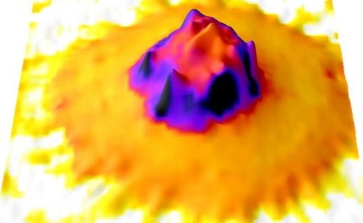
How do leukocytes "know" where to start working at a site of inflammation? The European MechaDynA (Multi-scale mechanics of dynamic leukocyte adhesion) project, supervised by Felix RICO (DyNaMo), uses revolutionary nanotools to discover the mechanical forces at play.
The immune system's ability to detect and respond to injury or infection begins with leukocytes, or white blood cells, patrolling vascular walls in search of inflammation. This process begins with a reduction in the speed of leukocytes as they pass through the bloodstream. These can then be activated, adhering firmly to the vascular wall and migrating to sites of injury or inflammation. Leukocyte adhesion to the vascular wall involves several steps, and exposes the cells to mechanical forces over a period of time ranging from a few microseconds to several minutes. Understanding the forces involved in the leukocyte adhesion cascade is key to deciphering the fundamental physics of immune response mechanisms.
Combined nanotools to measure force in leukocytesTo
fully understand the leukocyte adhesion process, the ERC-funded MechaDynA project aimed to develop a comprehensive approach that takes account of these time scales and spatial resolutions. The research team adapted two state-of-the-art nanotools, high-speed force spectroscopy (HS-FS) and acoustic force spectroscopy (AFS), to measure, directly on living cells, the mechanics of all cellular components involved over the widest range of time scales. Derived from atomic force microscopy, HS-FS uses ultra-small cantilevers to measure the mechanical properties and binding forces of individual molecules, such as proteins or DNA, on extremely short timescales of the order of microseconds. This approach has enabled researchers to explore the rapid events essential to leukocyte adhesion. AFS complements the time scales accessible by HS-FS. By using acoustic waves to trap and apply forces to hundreds of particles or cells suspended in parallel, it enables long-term measurements (several hours).
Experimentation on living
cellsOne of the main achievements of the project was the adaptation of HS-FS and AFS to work directly on living cells, enabling detailed exploration of leukocyte mechanics and adhesion. "By combining these tools with advanced optical microscopy, we were able to measure force on living cells with access to an unprecedented range of time scales," emphasizes MechaDynA principal investigator Felix Rico. Part of the project revealed that adhesion is linked to the mechanical stiffness of leukocyte cells. Interestingly, the stiffening seems to persist even after adhesion has been eliminated, highlighting a previously unknown aspect of cell mechanics. Overall, MechaDynA's results improve our understanding of the mechanical forces that influence biological processes.
Future
applicationsMechaDynA's success in adapting and applying HS-FS and AFS to the study of leukocytes marks an important turning point in biophysical research. The next steps in the project will be to extend these methods to other cell systems, such as circulating tumor cells, which resemble leukocytes moving through the bloodstream before metastasizing to distant organs. Exploring the mechanics of these cells could lead to breakthroughs in cancer diagnosis and treatment. In addition, MechaDynA's nanotools hold promise for the study of other biological systems, such as virus-cell interactions, which could advance our understanding of viral infections and contribute to the development of antiviral therapies. The possibility of patenting these nanotools underlines their innovative character and applicability across multiple disciplines, and opens up new avenues for tackling pressing biomedical challenges. In addition, the PyFMLab open-source software package developed as part of the project offers a standardized solution for the viscoelastic characterization of biological samples.
Link to CORDIS article: https://cordis.europa.eu/article/id/456256-the-forces-that-power-immune…